Procedures
EUS-Guided Biopsy (FNA and FNB) — How Samples Are Taken
There is a point in many pancreatic and lymph node investigations where imaging has gone as far as it can. The CT shows a mass. The EUS shows its size, its edges, and whether it has involved a blood vessel. What no scan can tell you is what the mass is actually made of — and until that is known, nobody can plan treatment.
That is what EUS-guided sampling is for.
Why sample through the stomach wall?
The pancreas is one of the least accessible organs in the body. It sits behind the stomach, in front of the great vessels, surrounded by structures you very much do not want a needle passing through. Historically, getting tissue meant either a needle through the abdominal wall under CT guidance, or an operation.
EUS changed the geometry. With the scope inside the stomach or duodenum, the needle has only a few millimetres to travel, and the ultrasound displays the entire needle path in real time — including the vessels to avoid. The gut wall then heals as it does after any endoscopic biopsy.
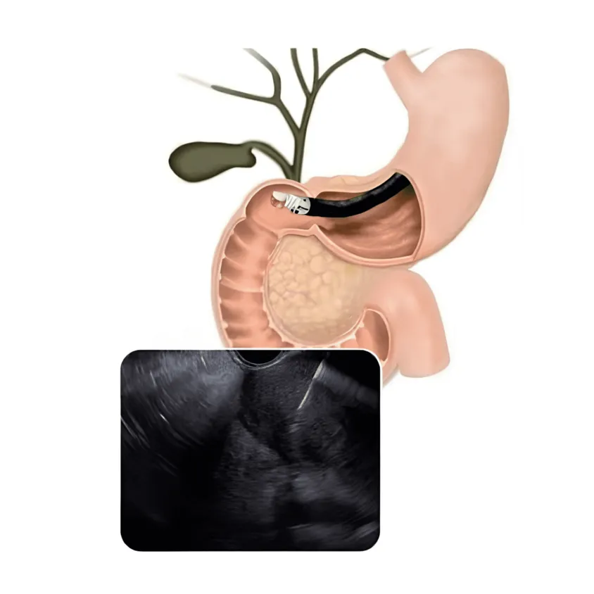
Figure 1: Anatomical view and the corresponding live sonographic tracking of needle deployment into the target. © Dr. Ali Taj
The same logic applies to lymph nodes alongside the oesophagus and stomach, to lesions in the left lobe of the liver, and to lumps arising within the gut wall itself.
Yeh kis liye hota hai? (What it is used to diagnose)
- Pancreatic masses and cysts: separating adenocarcinoma from an inflammatory mass or a benign cyst, which look similar on CT but are managed in completely different ways.
- Staging luminal cancers: establishing how deeply an oesophageal, gastric or rectal tumour has grown into the wall.
- Enlarged lymph nodes in the mediastinum or abdomen.
- Subepithelial lesions: a bulge seen at ordinary endoscopy where the question is what it arises from.
FNA or FNB — what is the difference?
| FNA (fine needle aspiration) | FNB (fine needle biopsy) | |
|---|---|---|
| What it collects | Loose cells | A small core of intact tissue |
| Read as | Cytology | Histology |
| Shows tissue architecture | No | Yes |
| Best for | Confirming a suspected carcinoma, cyst fluid | Lymphoma, autoimmune pancreatitis, tumours needing special stains |
| Passes usually needed | More | Fewer |
Practice has moved steadily towards FNB needles, because a core of tissue supports the immunohistochemistry that many diagnoses now depend on. Cytology alone can confirm that malignant cells are present, but frequently cannot say what kind — and “what kind” is what determines treatment.
The tuberculosis question
This deserves its own section, because it is where practice in Karachi departs from the assumptions built into the Western textbooks.
When a patient in Europe presents with enlarged mediastinal or peri-gastric lymph nodes and weight loss, the working diagnosis is malignancy. In Pakistan, tuberculosis sits alongside it as a genuinely competing possibility — and on CT the two can be indistinguishable. Getting it wrong is serious in both directions: months of anti-tuberculous therapy for what is actually a cancer, or an oncology work-up for treatable TB.
Needle sampling resolves it. Where TB is a real consideration, part of the sample goes for mycobacterial culture and molecular testing alongside the cytology, not merely for a look under the microscope. That request has to be made at the time of the procedure, which is why the clinical question needs settling before the needle goes in rather than after the report comes back.
What happens during sampling
Sampling is done during the same EUS session, under the same sedation. It is not a separate appointment.
Once the target is in view, the needle is advanced down the working channel of the scope and through the gut wall into the lesion, its path visible throughout. Several passes are usually made, each moving the needle back and forth within the lesion to collect material. The samples are then prepared and sent to pathology.

Figure 2: Acoustic mapping of a sub-mucosal lesion to guide safe fine-needle tracking. © Dr. Ali Taj
Adding sampling extends the procedure by roughly fifteen to thirty minutes. Everything else about the day (fasting, sedation, arranging someone to take you home) is the same as for a diagnostic EUS.
Risks specific to needle sampling
Diagnostic EUS on its own is very safe. Most of the small risk attached to EUS comes from the needle, which is exactly why sampling is not routine:
- Pancreatitis: when the needle passes through pancreatic tissue. The main one, and uncommon.
- Bleeding: usually minor and self-limiting, occasionally into a cyst.
- Infection: mainly relevant when sampling or draining cystic lesions, which is why antibiotics are given for those.
- A non-diagnostic sample: not a complication as such, but it happens, and it may mean repeating the procedure.
After an EUS with biopsy, seek urgent help if you develop:
- Severe abdominal pain, especially pain radiating to the back, that worsens over hours
- Fever or shaking chills
- Vomiting blood, or passing black tarry stools
- Light-headedness or fainting
Post-procedure pancreatitis declares itself as pain that escalates rather than eases. Early assessment matters.
Getting the results
Dr. Taj can describe what the images showed once you are awake. The tissue result takes several working days, because the sample has to be processed, stained and read — and if the first assessment is inconclusive, further stains add more time.
It is worth being prepared for the possibility that the answer comes back as “insufficient material”. It is frustrating, it is nobody’s fault, and it sometimes means a second attempt.
Karachi mein EUS-guided biopsy
Sampling yield depends heavily on the operator: how the target is approached, how many passes are made, and how the material is handled at the bedside.
Dr. Muhammad Ali Taj is a consultant gastroenterologist and interventional endoscopist in Karachi with 28+ years in practice and over 14,000 endoscopic procedures, including diagnostic and therapeutic EUS and ERCP. Qualifications: MBBS, MCPS, FCPS (Gastroenterology), MRCP Gastroenterology (UK), SCE (UK), ESEGH (Europe). His publications are listed on Google Scholar and ORCID.
He practises at Ziauddin Hospital Clifton, Hill Park General Hospital, Life Care Consultant Clinics, and Usman Memorial Hospital.
Aksar puchhe jaane wale sawaalat (FAQs)
Why do I need an EUS biopsy instead of a normal one? Because the target cannot be reached any other way. The pancreas sits deep behind the stomach, and lymph nodes near the oesophagus sit behind the chest wall. EUS puts the needle within millimetres of these structures through the gut wall, avoiding a surgical procedure or a needle passed through the abdominal skin.
What is the difference between FNA and FNB? FNA (fine needle aspiration) draws out individual cells, which a pathologist examines as cytology. FNB (fine needle biopsy) uses a needle designed to cut a small core of tissue, preserving how the cells are arranged. FNB is chosen when the diagnosis depends on that architecture, such as in lymphoma or some pancreatic tumours.
How accurate is EUS-guided biopsy? For solid pancreatic masses it is highly accurate in experienced hands, and it is the established first-line method of obtaining tissue. Accuracy depends on the operator, the number of needle passes, and how the sample is handled afterwards. A non-diagnostic result does happen, and the procedure occasionally has to be repeated.
Is EUS-FNA dangerous? Complications are uncommon but real. The main ones are bleeding, infection, and pancreatitis when the needle passes through pancreatic tissue. This is why sampling is only performed when the result will actually change management, rather than as a routine addition to every EUS.
How long do biopsy results take? Usually several working days. The sample goes to a pathologist for processing, staining and interpretation, and additional stains may be requested if the first look is inconclusive. Dr. Taj will tell you what the images showed on the day, but the tissue answer takes longer.
Can an EUS biopsy spread cancer? This concern comes up often. Needle-track seeding is documented but very rare with EUS-guided sampling, partly because the needle path is short and often runs through tissue that would be removed during any subsequent surgery. The risk of not knowing the diagnosis is far greater than this theoretical risk.
General information only, not a substitute for consultation. Whether tissue sampling is needed depends on your imaging and clinical picture.