Understanding Endoscopic Ultrasound (EUS)
This blog post aims to inform and guide patients about the EUS procedure and the expertise Dr. Ali brings to this advanced diagnostic and therapeutic technique.
What is Endoscopic Ultrasound (EUS)?
Endoscopic Ultrasound (EUS) is a minimally invasive procedure combining endoscopy and ultrasound to provide detailed images of the digestive tract and surrounding tissues and organs. This technique is essential for diagnosing and staging gastrointestinal cancers, evaluating chronic pancreatitis, and assessing other conditions like gallstones.
The term EUS breaks down to :
- Endoscopic (Endoscope): Refers to the use of a long, thin, flexible tube equipped with a light source and a high-definition camera, to safely navigate your digestive tract.
- Ultrasound: Denotes the high-frequency sound wave technology utilized via a specialized acoustic transducer built directly into the tip of the endoscope.
- Sonography (Scanning): Represents the deep tissue imaging produced as these sound waves echo off internal organs, tissues, and blood vessels, mapping out areas that standard visual endoscopy cannot see.
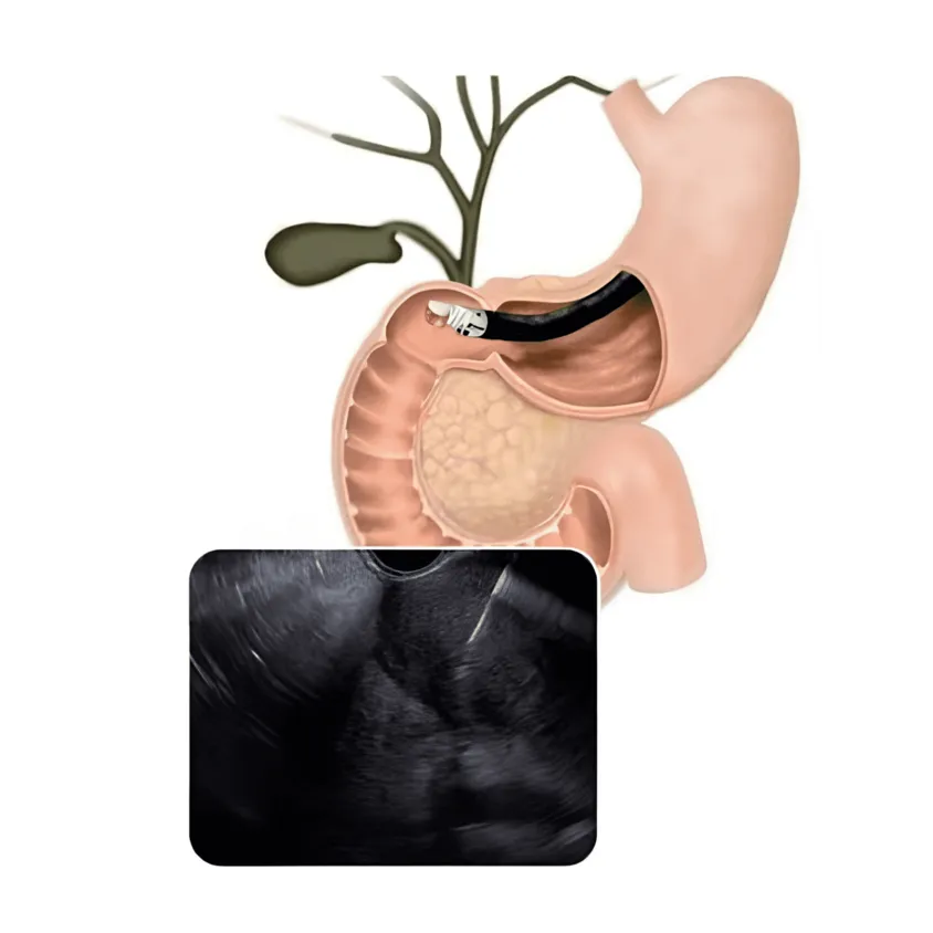
Figure 1: Simplified schematic showing acoustic wave emission from an internal endoscopic probe. © Dr. Ali Taj
Types of EUS tests
Endoscopic Ultrasound procedures are clinically categorized based on the specific section of the gastrointestinal tract undergoing evaluation. The two variations of this advanced diagnostic test include:
1. Upper EUS Procedure
An Upper EUS involves passing the specialized echoendoscope through the upper digestive tract. This diagnostic method combines standard upper endoscopy techniques with high-frequency scanning to examine:
- Upper Luminal Linings: The intricate tissue layers of the esophagus, stomach, and the initial portion of the small intestine.
- Hepatobiliary and Solid Organs: High resolution views of adjacent structures, including the pancreas, gallbladder, and bile ducts.
- Vascular and Lymphatic Pathways: Close assessment of regional lymph nodes, blood vessels, cysts, and abnormal tumors.
2. Lower EUS Procedure
A Lower EUS focuses exclusively on evaluating the structures of your digestive tract, with the echoendoscope introduced retrogradely. This specialized test maps out:
- Lower Intestinal Anatomy: The delicate mucosal and muscle linings of the large intestine, specifically targeting the rectum and colon.
- Anorectal Musculature: The structural integrity of the anal sphincter (the essential muscles surrounding the anus).
Indications for EUS
EUS is typically indicated for:
- Diagnosing and Staging GI Cancers: EUS provides critical information for assessing the stage of cancers in the GI tract, pancreas, liver, and other areas.
- Evaluating Chronic Pancreatitis and Pancreatic Cysts: It helps in assessing the severity of chronic pancreatitis and nature of pancreatic cysts.
- Investigating Mediastinal Diseases: EUS is useful in evaluating diseases in the mediastinum, like lymph node enlargement.
- Guiding Fine-needle Aspiration (FNA): For collecting tissue samples from areas that are hard to reach with traditional endoscopy.
Preparing for EUS
Before the Procedure
- Fasting: Patients are typically required to fast for at least 6 hours before the procedure.
- Medication Adjustments: Inform your doctor about all medications, especially blood thinners, as adjustments may be necessary.
- Health Conditions: Disclose any health conditions, allergies, or prior surgeries to your doctor.
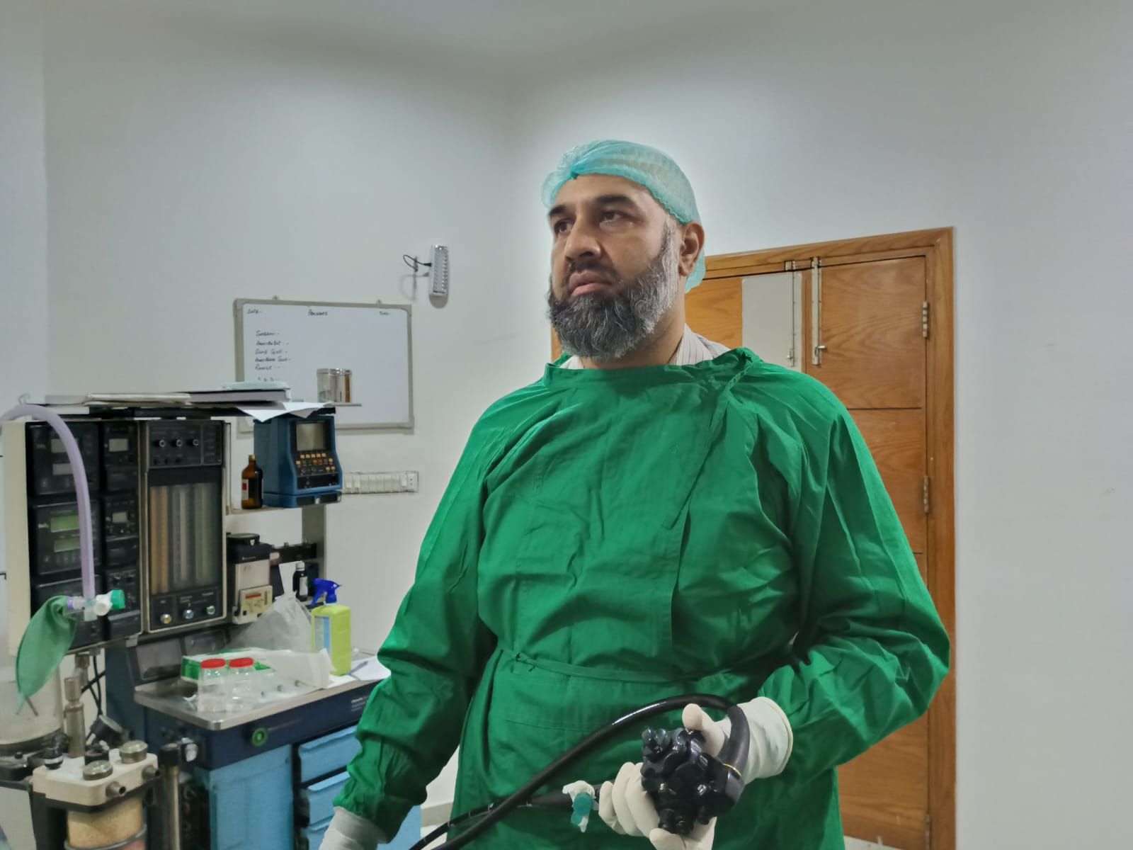
Dr. M. Ali Taj preparing advanced therapeutic tools for an EUS. © Dr. M. Ali Taj
During the Procedure
- Anesthesia: For comfort and to eliminate anxiety, the clinical team administers intravenous anesthesia before the procedure begins.
- Scope Insertion and Breathing: Dr. Ali gently guides the flexible ultrasound endoscope through your mouth, passing smoothly down the esophagus and stomach until it reaches the small intestine.
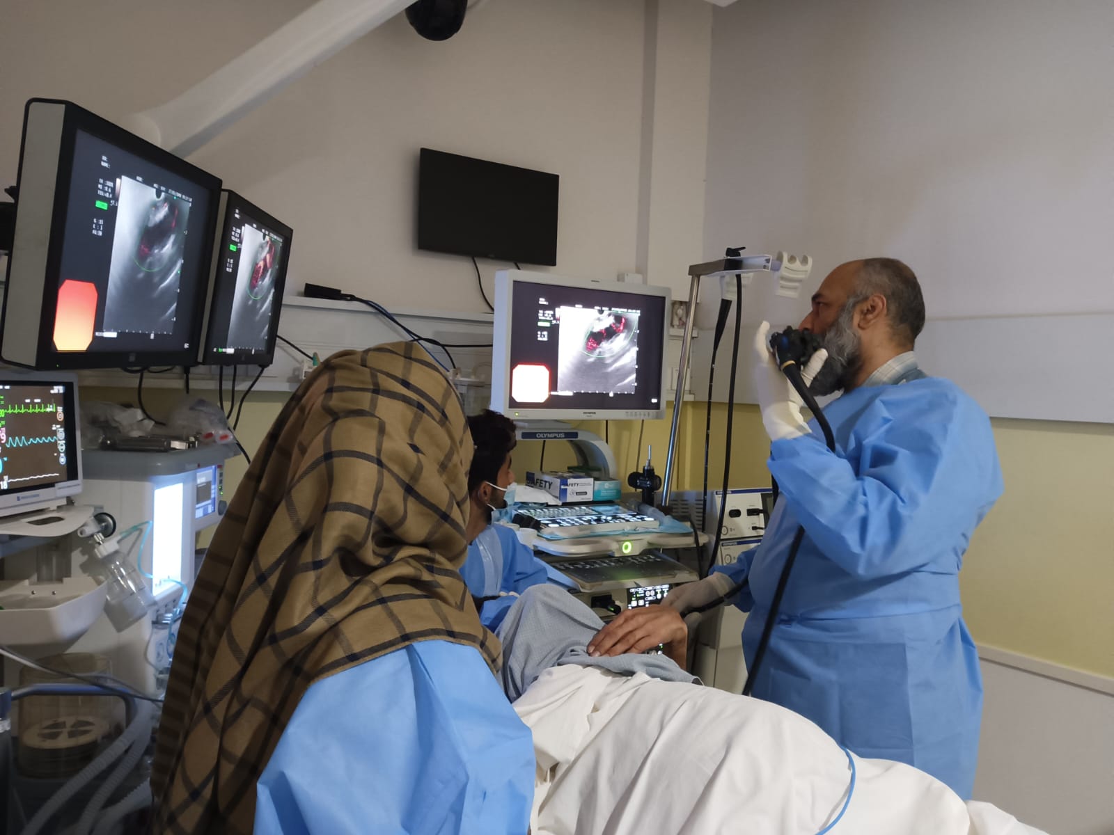
- Real-Time Monitoring: As the scope navigates your anatomy, the clinical team monitors live, high-resolution ultrasound feeds on a dedicated display screen. This allows for an immediate and highly detailed visual examination of your upper digestive tract, neighboring organs, cysts, or structural tumors.
- Targeted Tissue Sampling: If a specific lesion or abnormality is identified that warrants further laboratory evaluation, Dr. Ali uses the live sonographic guide to thread a micro-fine needle directly through the internal channel of the endoscope to securely extract a precise sample of tissue or fluid.
- Procedural Duration: Depending on the anatomical complexity of the target area and whether active fine-needle tissue collection is required, the entire procedure usually takes between 30 to 90 minutes to complete.
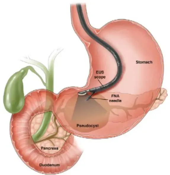
Detailed illustration of a stomach with an endoscopic ultrasound (EUS) scope positioned to evaluate internal GI tract layers and adjacent structures. © Dr. M. Ali Taj
Potential Risks or Complications
While EUS is generally safe, it’s important to be aware of potential risks, including:
- Mild Discomfort: Some patients may experience throat soreness or mild abdominal discomfort post-procedure.
- Infection: Rarely, infections can occur, particularly in procedures involving needle aspiration.
- Bleeding: This is a rare complication, usually associated with tissue sampling.
- Pancreatitis: In some cases, EUS can trigger pancreatitis, especially after interventions on the pancreas.
Post-Procedure Care
- Observation: Patients are monitored for a short period after sedation wears off.
- Transportation: Due to sedation, arrange for someone to drive you home.
- Follow-Up: Adhere to any specific post-procedure instructions given by your healthcare provider.
Schedule an Appointment
Learn More about Dr. Ali’s Services
Note: This content is designed for informational purposes only and should not replace professional medical advice. Always consult your doctor for personalized care.