Endoscopic Ultrasonography-Guided Fine-Needle Aspiration: Best Practices and Techniques
Introduction to Endoscopic Ultrasonography-Guided Fine-Needle Aspiration
Endoscopic Ultrasonography-Guided Fine-Needle Aspiration (EUS-FNA) is a pivotal diagnostic procedure in gastroenterology, offering a minimally invasive method to obtain tissue samples from lesions within or adjacent to the gastrointestinal tract. Mastery of EUS-FNA technique is critical for enhancing diagnostic yield while ensuring patient safety.
Essential Techniques in EUS-FNA
Keeping the Needle in the Visual Plane
The foremost principle in EUS-FNA is maintaining the needle within the visual plane at all times. This approach minimizes the risk of complications by allowing the endosonographer to accurately guide the needle into the target lesion, ensuring precise sampling and reducing the likelihood of damaging surrounding structures.
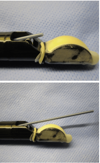
Figure 20.1: Structural tracking of the FNA needle path, illustrating the visual cross-section profile under real-time ultrasound profiling. © Dr. Ali Taj
Avoiding Excessive Force
When navigating the needle sheath past acute bends in the endoscope tip, it is imperative to avoid using excessive force. Gentle manipulation prevents damage to the endoscope and minimizes patient discomfort, contributing to the overall success and safety of the procedure.
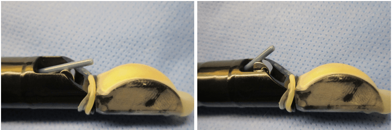
Figure 20.2: Side-by-side tip comparison demonstrating proper elevator mechanics to modify the needle insertion angle smoothly without damaging the inner channel sheath. © Dr. Ali Taj
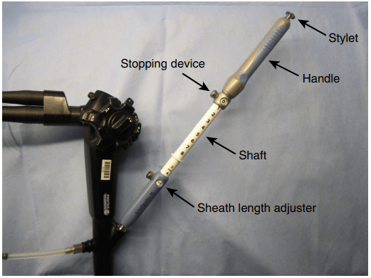
Reevaluating the Use of the Stylet
Contrary to common practice, using a stylet during EUS-FNA does not increase the yield of tissue samples and can be more cumbersome. Simplifying the procedure by omitting the stylet may enhance efficiency without compromising sample quality, streamlining the process for both the practitioner and patient.
Selecting the Optimal Needle Gauge
The choice of needle gauge significantly influences diagnostic yield, particularly when sampling pancreatic lesions. Twenty-five-gauge (25-G) needles are shown to provide a superior diagnostic yield compared to 22-gauge (22-G) needles, offering a finer balance between sample adequacy and ease of needle manipulation.
Best Practices for Cyst Aspiration
When aspirating a cyst, the endosonographer should aim to fully aspirate all fluid to minimize the risk of recurrence and infection. Making only one pass with the needle, using prophylactic antibiotics, and avoiding attempts to perform aspiration cytology from the cyst wall are recommended practices to enhance patient outcomes and reduce complications.
Conclusion
Endoscopic Ultrasonography-Guided Fine-Needle Aspiration is a complex yet crucial procedure in the diagnosis of gastrointestinal lesions. Adhering to best practices and refined techniques, such as maintaining visual control of the needle, selecting the appropriate needle gauge, and adopting a judicious approach to cyst aspiration, can significantly improve diagnostic accuracy and patient safety. By continually evolving our understanding and application of EUS-FNA, we can further the field of gastroenterology and offer better care to our patients.
Disclaimer: This content is for informational purposes only and should not be considered as medical advice. Always consult a healthcare professional for personal medical advice.
Learn More About Our Gastroenterology Services
Contact Us for More Information