
Advancements in Endoscopic Ultrasonography: Focusing on the Esophagus and Mediastinum
This article explores the significant role of Endoscopic Ultrasonography (EUS) in evaluating the esophagus and mediastinum, highlighting its precision and correlation with computed tomography (CT) imaging.
Endoscopic Ultrasonography in the Esophagus and Mediastinum
Endoscopic Ultrasonography (EUS) has become a pivotal tool in assessing conditions related to the esophagus and mediastinum. Its ability to provide detailed images has made it an indispensable method in gastroenterology and thoracic medicine.
Precision Imaging in the Mediastinum
EUS offers a straightforward approach to evaluating the mediastinum. The images obtained by the radial scanning instrument in EUS correlate closely with those of CT scans, providing a high level of precision and detail.
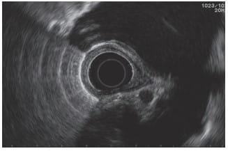
Radial EUS cross-section mapping the mediastinal vascular structures and corresponding anatomical planes. © Dr. Ali Taj
Interpretation of Linear Images
Linear images in EUS, though more challenging to interpret, can yield significant insights when examined systematically. Mastery of this technique, particularly using a linear array echoendoscope, is crucial for a successful examination of the mediastinum.
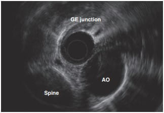
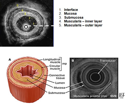
Linear array EUS tracing the structural layers of the esophageal wall during a longitudinal sweep. © Dr. Ali Taj
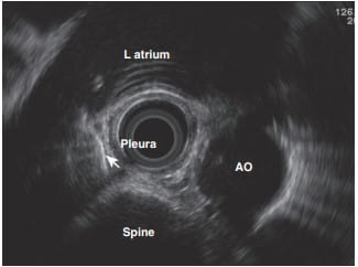
Linear EUS visualization assessing posterior mediastinal lymph node structures and surrounding tissue. © Dr. Ali taj
Systematic Approach for Effective Evaluation
To effectively use EUS in the mediastinum, a systematic approach is essential, especially when working with linear array echoendoscopes. This method ensures comprehensive and accurate assessments.
- Structured Examination: Following a structured protocol ensures that all relevant areas are thoroughly examined.
- Comparative Analysis with CT: Correlating EUS findings with CT scans enhances the understanding and interpretation of images.
- Expert Interpretation: Due to the complexity of linear images, expert interpretation is necessary to accurately diagnose and assess conditions.
The Clinical Impact of EUS
EUS has revolutionized the way the esophagus and mediastinum are evaluated. Its high-resolution imaging capability allows for early detection and accurate staging of various conditions, leading to improved patient outcomes.
Conclusion
Endoscopic Ultrasonography has solidified its role as a critical diagnostic tool in assessing esophageal and mediastinal conditions. Its ability to provide detailed and precise images, comparable to CT scans, makes it an invaluable asset in modern gastroenterology and thoracic medicine.
Disclaimer: This content is for educational purposes only and should not be considered a substitute for professional medical advice. Always consult a healthcare provider for accurate diagnosis and treatment.
Contact Us for More Information
Explore Our Endosonography Services
References
- Findings of Endoscopic US and CT of Esophageal Disease: A Complementary Approach
- Endoscopic ultrasound in the diagnosis of mediastinal diseases and tissue acquisition
- Revised ESTS guidelines for preoperative mediastinal lymph node staging for non-small-cell lung cancer
- Mediastinal Nodal Staging Performance of Combined Endobronchial and Esophageal Endosonography in Lung Cancer Cases